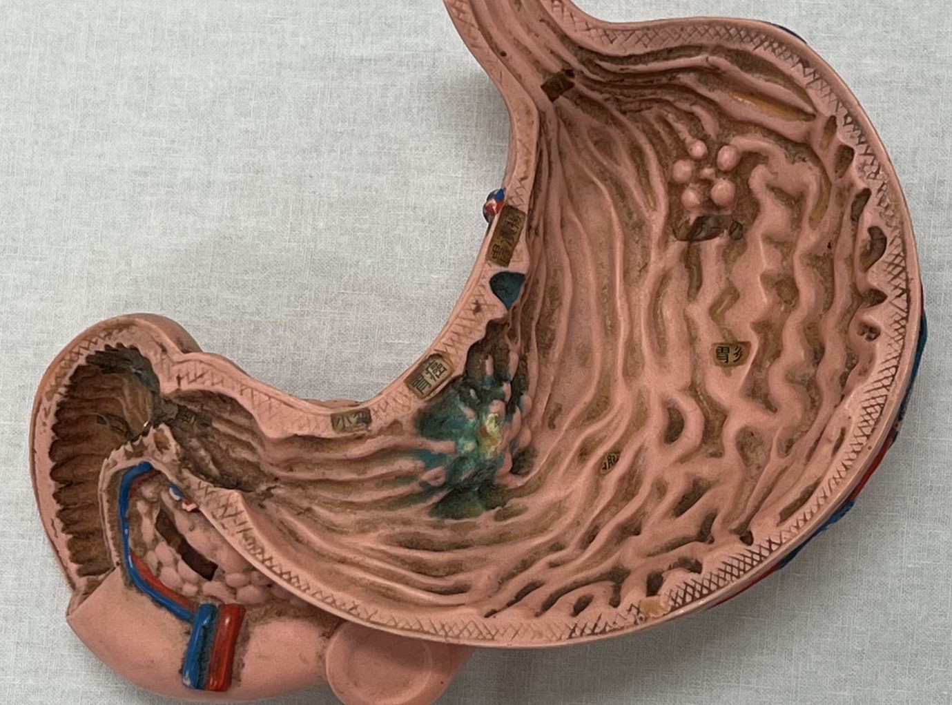
What Is a Gastroscopy and Why Is It Performed?

Gastroscopy, also known as upper gastrointestinal (GI) endoscopy or esophagogastroduodenoscopy (EGD), is an examination that allows the doctor to directly view the inside of the esophagus, stomach, and duodenum using a thin, flexible endoscope equipped with a small camera.
The examination provides detailed images of the lining of the upper gastrointestinal tract, allowing the doctor to identify abnormalities and changes that may require further evaluation or treatment.
One of the important benefits of gastroscopy is its ability to detect certain
conditions at an early stage, sometimes before they cause significant symptoms.
Early detection can help guide appropriate treatment and follow-up.
Experienced Specialist and Advanced Fujifilm Endoscopy Technology
Gastroscopy at Horii Clinic is performed by an experienced specialist in gastrointestinal endoscopy.
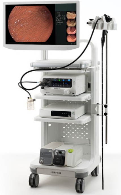
Our clinic uses an advanced Fujifilm endoscopy system with high-resolution imaging and image-enhancement technologies. These features help the doctor examine the gastrointestinal lining in detail and identify subtle changes that may require further evaluation.
When appropriate, testing for Helicobacter pylori (H. pylori), a major cause of gastritis and peptic ulcers, may also be performed
during the examination, including by taking a small tissue sample (biopsy).
Our Approach to Gastroscopy
Experienced Endoscopy Specialist
Gastroscopy is performed by an experienced specialist in gastrointestinal endoscopy.
Careful, Detailed Examination
We allow at least 7 minutes for careful observation during the examination, helping the doctor assess the gastrointestinal lining in detail.
Same-Day Examination When Available
Depending on your symptoms, medical needs, and appointment availability, a same-day gastroscopy may be possible.
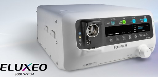
Advanced Fujifilm Endoscopy System
Our clinic uses the Fujifilm EP-8000 / EG-840N endoscopy system for high-resolution examination of the upper gastrointestinal tract.
BLI and LCI Image-Enhancement Technologies
Blue Light Imaging (BLI) and Linked Color Imaging (LCI) provide enhanced
visualization of the gastrointestinal lining, helping the doctor identify
and evaluate subtle abnormalities.



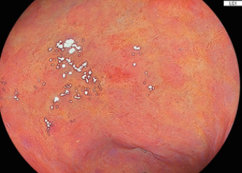
Advanced Imaging for Detailed Examination
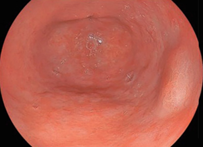
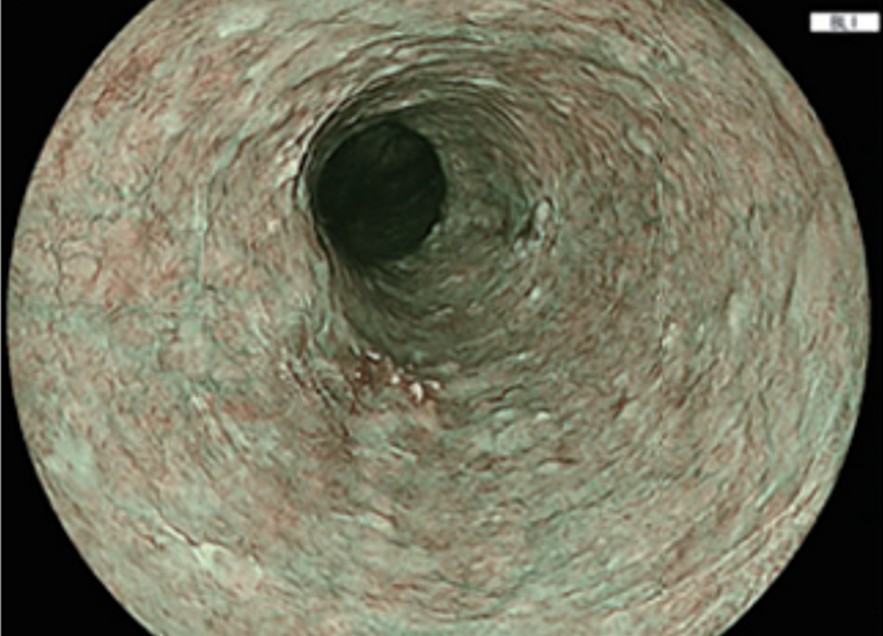
Our clinic uses a high-resolution endoscopy system designed to provide clear, detailed images of the gastrointestinal lining. This helps the doctor identify subtle changes that may require closer evaluation.
4-LED Multi-Light Technology
Our Fujifilm endoscopy system uses 4-LED Multi-Light Technology, which combines different wavelengths of light to enhance visualization of the gastrointestinal lining.
By providing different imaging modes, this technology can help the doctor identify and evaluate subtle changes in the mucosa, including abnormalities that may be associated with precancerous or early-stage lesions.
Enhanced visualization can support earlier detection and help the doctor
determine whether further evaluation, follow-up, or treatment is appropriate.
Care and Support Throughout Your Examination
Clear Explanation and Follow-Up
Our care does not end when the examination is completed. After your gastroscopy, the doctor will explain the findings in clear, easy-to-understand language and provide you with a detailed examination report.
When further evaluation or treatment is needed, we will discuss the appropriate
next steps with you and, when necessary, coordinate care with a gastroenterology
department or specialist at another medical institution.
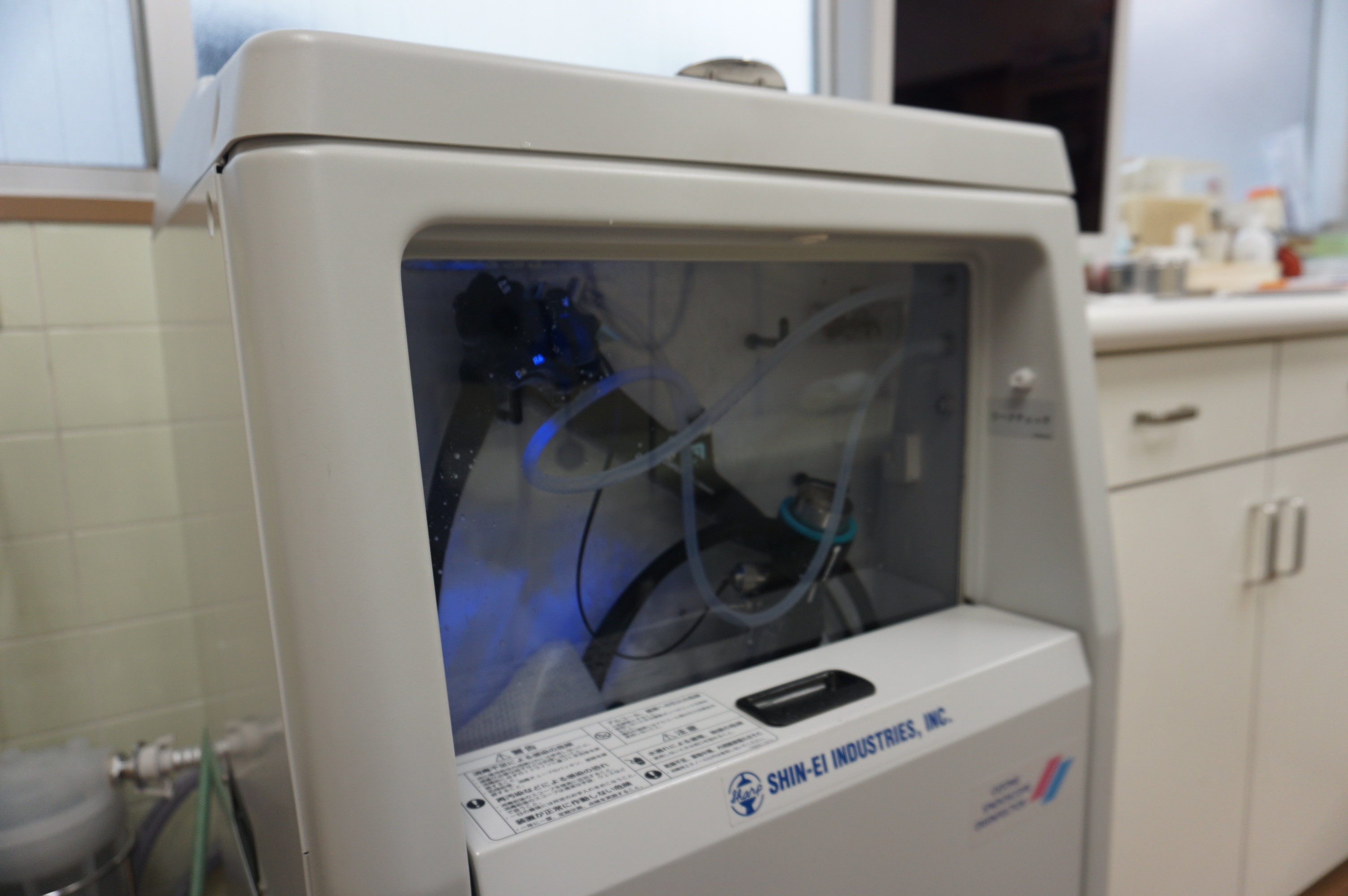
Endoscope Cleaning and Disinfection
Patient safety and infection prevention are important parts of endoscopic care.
After use, endoscopes are cleaned and disinfected using an automated endoscope
reprocessor. This process helps maintain appropriate hygiene standards
and reduce the risk of infection associated with endoscopic procedures.
Comfortable, Patient-Centered Endoscopy Care
We aim to make your gastroscopy as comfortable as possible in a calm, well-equipped examination environment.
When appropriate, medications such as sedation or local anesthesia may be used to help reduce discomfort during the examination. The doctor will consider your individual health and medical needs when determining the appropriate approach.
If an abnormality is identified during gastroscopy, additional procedures such as a biopsy may be performed during the same examination when appropriate.
What Can Gastroscopy Detect?

During gastroscopy, the doctor examines the lining of the esophagus, stomach,
and duodenum in real time using high-resolution images.
Gastroscopy can help identify and evaluate a range of abnormalities, including:
Changes in the gastrointestinal lining
The doctor assesses the color, surface appearance, and folds of the lining for changes that may indicate an abnormality.
Inflammation, atrophy, and vascular changes
Gastroscopy can help identify signs of gastritis, thinning or loss of the normal gastric lining (atrophy), and abnormalities in the appearance of blood vessels.
Erosions, ulcers, and tears
The examination can identify damage to the gastrointestinal lining, including erosions, gastric or duodenal ulcers, and mucosal tears that may be associated with pain or gastrointestinal bleeding.
Polyps and tumors
High-resolution endoscopy allows the doctor to identify and evaluate polyps and other lesions, including abnormalities that may require further examination, biopsy, or treatment. High-resolution endoscopy can also help identify lesions beneath the surface of the lining, including subepithelial lesions.
What Information Can Gastroscopy Provide?

Gastroscopy provides detailed information that can help the doctor evaluate
conditions affecting the esophagus, stomach, and duodenum.
It can help the doctor:
Detect disease at an early stage
Gastroscopy can help identify abnormalities of the esophagus, stomach, and duodenum at an early stage.
Evaluate the extent and characteristics of abnormalities
When a lesion is identified, gastroscopy can provide information about its location, extent, appearance, and other important features.
Distinguish between conditions with similar symptoms or findings
Endoscopic findings can help the doctor differentiate between conditions that may have similar symptoms or clinical features.
Identify signs of complications
Gastroscopy can help identify complications such as bleeding from gastric
or duodenal ulcers and varices in the esophagus or stomach. It can also
help assess findings associated with deep ulcers that may require further
evaluation or treatment.
Advantages of Gastroscopy
Unlike imaging examinations such as a barium study, gastroscopy allows the doctor to directly examine the lining of the esophagus, stomach, and duodenum.
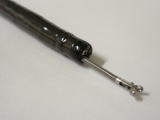
Another important advantage is that diagnostic and, when appropriate, therapeutic procedures can be performed during the same examination. Small instruments can be passed through a dedicated channel in the endoscope, allowing the doctor to perform procedures such as:
Biopsy
A small tissue sample can be taken from an abnormal area and sent for pathological examination, including evaluation for abnormal or cancerous cells.
Therapeutic Procedures
When appropriate, endoscopic treatment may be performed during the examination,
such as controlling gastrointestinal bleeding or removing a foreign object.
When May Gastroscopy Be Recommended?

Gastroscopy may be recommended when certain conditions affecting the upper gastrointestinal tract are suspected or require further evaluation.
These may include:
Gastritis and duodenitis
Inflammation of the lining of the stomach or duodenum.
Erosions and peptic ulcers
Damage to the lining of the stomach or duodenum, including gastric and duodenal ulcers.
Gastroesophageal reflux disease (GERD)
A condition in which stomach contents flow back into the esophagus and may cause symptoms such as heartburn.
Esophageal diverticula
Pouch-like areas that develop in the wall of the esophagus.
Gastric intestinal metaplasia
Changes in the stomach lining that may be associated with an increased risk of gastric cancer.
Crohn’s disease
A chronic inflammatory condition of the gastrointestinal tract.
Polyps and tumors
Gastroscopy can help identify and evaluate benign or malignant lesions
in the esophagus, stomach, and duodenum.
When Should You Consider a Gastroscopy?

A gastroscopy may be recommended if you have persistent or unexplained symptoms affecting the upper gastrointestinal tract.
These may include:
Nausea, vomiting, frequent belching, or persistent bloating
Heartburn or difficulty swallowing
Persistent or recurrent abdominal pain
Vomiting blood or passing black, tarry stools
Unexplained weight loss or loss of appetite
Changes in taste or a persistent unpleasant taste in the mouth
Unexplained anemia
Gastroscopy may also be recommended for routine screening or follow-up,
particularly if you have previously been diagnosed with a gastrointestinal
condition or have undergone treatment that requires ongoing monitoring.
Is Gastroscopy Safe?

Gastroscopy is generally a safe and well-established examination that can be performed in many patients.
Before the examination, the doctor will review your medical history, current health, medications, and any other factors that may require special precautions.
Depending on your individual condition, the examination may need to be
modified, postponed, or performed with additional precautions. If you have
any concerns about whether gastroscopy is appropriate for you, please discuss
them with the doctor before the examination.
Preparing for Your Gastroscopy
Proper preparation is important to ensure that the doctor can examine your upper gastrointestinal tract clearly and safely. Please follow the instructions provided by the clinic before your examination.
Eating and Drinking

Gastroscopy must be performed on an empty stomach. Please do not eat for 10–12 hours before your examination.
If your examination is scheduled for the evening, you may have a light breakfast in the morning. After that, please do not eat until your examination has been completed.
Please follow the clinic's instructions regarding what you may drink before the examination.
Medical Conditions and Allergies

Before your examination, please tell the doctor about:
• Any chronic or ongoing medical conditions
• Any allergies, including allergies to medications
• Any medications you are currently taking
• Any recent examination or test results that may be relevant
If you have recent medical records or test results, please bring them with you.
What to Bring

Please bring:
• A valid form of identification
• Your Japanese health insurance card or other health insurance information,
if applicable
• Your residence card or passport, if applicable
• A referral letter, if you have one
• Any relevant medical records or recent test result
What to Expect on the Day of Your Gastroscopy

1. Appointment
Gastroscopy is performed by appointment. Please arrange your examination in advance.
2. Check-In
When you arrive at the clinic, our staff will confirm the necessary information and documents before your examination.
Please bring the identification, health insurance information, and any relevant medical records or referral letter described in the preparation instructions above.
3. Gastroscopy
Once the necessary preparation has been completed, the doctor will begin the examination.
The observation itself usually takes approximately 5–7 minutes. If a biopsy
or another procedure is required during the examination, it may take longer.
Transoral or Transnasal Gastroscopy
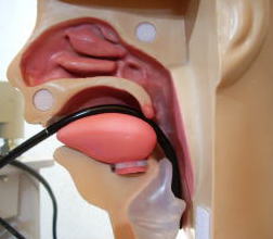
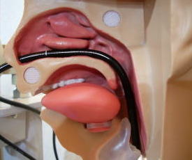
At Horii Clinic, gastroscopy can be performed either through the mouth (transoral gastroscopy) or through the nose (transnasal gastroscopy).
The doctor will discuss the available options with you before the examination so that the most appropriate method can be selected.
Transoral Gastroscopy
For a transoral gastroscopy, local anesthesia is applied to the throat before the examination to help reduce discomfort.
A mouthpiece is then placed between your teeth to protect both your mouth and the endoscope during the examination.
Transnasal Gastroscopy
For a transnasal gastroscopy, a local anesthetic gel containing lidocaine is applied inside the nose before the examination.
The endoscope is then gently inserted through the nose and advanced into
the upper gastrointestinal tract.
During and After Your Gastroscopy

During the Examination
You will lie on your left side on the examination bed.
The doctor will insert the endoscope through your mouth or nose, depending on the method selected, and carefully examine the esophagus, stomach, and duodenum while viewing high-resolution images on a monitor.
If an abnormal area is identified, a small tissue sample (biopsy) may be taken for pathological examination when appropriate. Other procedures, such as controlling gastrointestinal bleeding or removing a foreign object, may also be performed when necessary.
After the Examination
After the examination, the doctor will review the findings and explain the results to you. Your examination report and relevant endoscopic images will also be provided.
The results are usually available approximately 15–20 minutes after the examination.
Because local anesthesia is used during gastroscopy, please do not eat
or drink for approximately 30–60 minutes after the examination, or until the effects of the anesthesia have worn off.
How to Request an Appointment
You can request an appointment for a gastroscopy at Horii Clinic in either of the following ways:
Submit an appointment request through the online form on our website
Speak to our reception staff when visiting the clinic
For information about consultation, examination, and procedure fees, please see our Fees and Pricing page.
Please note that all examinations and procedures require a medical consultation
with the doctor beforehand.